Endometriosis ~ Abdominal Pain ~ Endo ~ Scar Tissue ~ Adhesions ~ Infertility ~ Hysterectomy
Showing posts with label Cul-de-sac Obliteration. Show all posts

Friday, May 02, 2014

First Harry Reich Award presented at EFA’s Blossom Ball

First Harry Reich Award presented at EFA’s Blossom Ball 
8 MARCH 2014
Professors Linda Giudice and Liselotte Mettler were honoured at EFA’s 6th Annual Blossom Ball with the inaugural Harry Reich Award.

Yesterday evening, the Endometriosis Foundation of America (EFA) awarded the inaugural Harry Reich Award to recognise two extraordinary healthcare professionals, Professors Linda Giudice and Liselotte Mettler, for the difference these two women have made in the lives of other women through their practice, research, and advocacy.

Tamer Seckin presenting Linda Giudice with her Award at the Blossom Ball
In announcing these two awards, co-founder of EFA, Dr Tamer Seckin, emphasised how the achievements of both Linda Giudice and Liselotte Mettler are an inspiration and encouragement for women (with and without endometriosis) to take charge of their health!
Professor Giudice is a biochemist, gynaecologist, and reproductive endocrinologist with a specific clinical interest in endometriosis, infertility, assisted reproduction, and implantation and ovulatory disorders.
Her research focuses on environmental impacts on reproductive health, steroid hormone signalling in human endometrium, endometrial-placental interactions, endometrium as a mucosal tissue, and translational applications of human embryonic and endometrial stem cells.  She is distinguished professor and chair of the Department of Reproductive Sciences at the University of California San Francisco, immediate past-president of the ASRM, president-elect of the World Endometriosis Society, vice-president of the World Endometriosis Research Foundation, and a member of the Institute of Medicine of the National Academy of Sciences.
Tamer Seckin presenting Liselotte Mettler with her Award at the Blossom Ball
Professor Emeritus Mettler has specialised in reproductive medicine, gynaecological endoscopy, endometriosis, and gynaecological endocrinology since 1981 when she became deputy director of the Department of Obstetrics and Gynaecology at the University of Kiel.  Following her retirement she remains an honorary patron of the Kiel School of Gynaecological Endoscopy and Reproductive Medicine, where she still teaches up to a dozen international training courses each year.
She is also visiting professor to the German Medical Center and the Dubai Healthcare City.  Professor Mettler is a former board member and current ambassador of the World Endometriosis Society, a board member of the German Foundation for Endometriosis Research (SEF), and the General Secretary of the International Academy of Human Reproduction.

The Harry Reich Award

The Harry Reich Award is awarded by EFA to recognise extraordinary healthcare professionals, who are making a difference in women’s lives (and their families) by their practice, research, and advocacy.

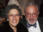
Harry Reich with honouree Linda Giudice at the Blossom Ball
The award is named for Dr Harry Reich, a pioneer in the field of laparoscopic surgery, who performed many “firsts”, including: the first laparoscopic hysterectomy, the first pelvic lymphadenectomy for cancer, and the first excision of cul-de-sac endometriosis that included rectal resection.
Dr Reich, who has operated in more than 60 countries, is an honorary professor in Russia and Romania, as well as a Fellow (ad eundem) of the Royal College of Obstetricians and Gynaecologists in the UK for his pioneering work in the field of endoscopy.  Though retired, Dr Reich remains actively involved in many medical organisations and serves as a reviewer for multiple scientific journals.
Please visit Endometriosis.org for more news about endo.

Dr. Drew doesn’t understand your “garbage” diagnosis ~ Endometriosis and Interstitial Cystitis

Grrrrrrrrrr. All the work we do for awareness and then this schmuck comes along...

The "Loveline" cohost draws outrage for his comments on endometriosis


Dr. Drew Pinsky is a board certified internist and assistant clinical professor of psychiatry at USC, but most of us know him better as radio and cable television’s most tireless ambulance chaser. Over the years, he’s doled out a variety of less than sound and/or helpful ideas. But when he speculated recently to a “Loveline” caller about his fiancée’s endometriosis, he definitively made the case against getting medical advice from flippant talk show hosts.
As Erin Gloria Ryan first noted on Jezebel, Pinsky was fielding a call from a man who was concerned about his girlfriend’s “multitude of conditions,” including endometriosis, interstitial cystitis, lactose intolerance and what he described as “no stomach lining.” The good doctor quickly interjected before the listener could even pose his question, explaining, “These are what we call sort of functional disorders. Everything you mentioned, everything you mentioned, are things that actually aren’t discernibly pathological. They’re what we call ‘garbage bag diagnoses,’ when you can’t think of anything else, you go, ‘Eh, it’s that.’ So, it then makes me question why is she so somatically preoccupied that she’s visiting doctors all the time with pains and urinary symptoms and pelvic symptoms, and then that makes me wonder, was she sexually abused growing up?”
Though the caller did acknowledge that his fiancée had in fact survived abuse, let’s take a step back here and observe that he never said the woman was “preoccupied” and “visiting doctors all the time” – on the contrary, he said she almost always “refuses” to go to doctors, even when she’s “in so much pain.” But Dr. Drew had a handy explanation, stating, “Trust me, she saw lots of doctors before you.” He then went on to explain why her early abuse was causing her problems now. “When people have unexplained pain, pelvic pain, it’s called somatoform dissociation,” he said, “and the only way her body, which was suffering during those early experiences, can tell its tale of woe is with pain. And she really needs to see a trauma specialist, not a urologist. Know what I’m saying?” This was immediately segued with Pinksy’s colleague joking that an additional way someone could have unexplained pelvic pain was by having sex with the show’s guest, Alan Thicke. Charming.
~~~~~~~~~~~~~~~~~~~~~~~~~~~~~~~~~~~~~~~~~
Here is the link to the CNN contact form for Dr. Drew's show...also what I wrote. Please give them a piece of your mind. IC folks....go get him!
Dr Drews comments on endometriosis are outrageous. He is not educated in a disease that strikes more than one in ten women! Find another doctor! Chronic stage 4 endo with severe adhesions has almost killed me twice. It is real, incurable and I hope you devote air some time in spreading awareness about endometriosis to counteract  the sad stereotype that has made the burden of this horrible disease even harder. 


Thursday, April 17, 2014

Safety and Effect on Quality of Life of Laparoscopic Douglasectomy with Radical Excision for Deeply Infiltrating Endometriosis in the Cul-de-Sac.

 2014 Mar;24(3):165-70. doi: 10.1089/lap.2013.0270.

Safety and Effect on Quality of Life of Laparoscopic Douglasectomy with Radical Excision for Deeply Infiltrating Endometriosis in the Cul-de-Sac.

Abstract

Abstract Objective: To evaluate the safety and effects on health-related quality of life (HRQOL) of radical excision of deeply infiltrating endometriosis (DIE) in the cul-de-sac. Patients and Methods: This study included 390 patients with pathologically proven DIE in the cul-de-sacwho underwent laparoscopic surgery between January 2000 and December 2011. The preoperative and postoperative visual analog scale (VAS) pain scores and HRQOL data from the 36-item Short Form (SF-36) questionnaire were recorded in 343 patients. Surgical outcomes, complications, and HRQOL were compared between patients who underwent hysterectomy and those who did not. Data were analyzed using the paired t test, Student's t test, and Pearson's correlation analysis. Results: In the group overall, the VAS pain score and all SF-36 scales improved after surgery. In patients who underwent hysterectomy, all SF-36 scales improved except physical functioning, role-physical, general health, and vitality. In patients who did not undergo hysterectomy, all SF-36 scales improved except general health. There were significant associations between gonadotropin-releasing hormone agonist therapy and SF-36 mental health, SF-36 mental component summary, and oral medication use. The VAS pain score and SF-36 body pain score showed the greatest improvement, and the SF-36 general health score showed the least improvement. Patients who underwent hysterectomy had more severe disease and poorer surgical outcomes than those who did not undergo hysterectomy. Conclusions: Laparoscopic radical excision of DIE in the cul-de-sac is safe and significantly improves HRQOL, especially in terms of pain. The severity of endometriosis may affect the degree of improvement in HRQOL scores.
PMID:
 
24625348
 
[PubMed - in process]

Saturday, March 08, 2014

MIT bioengineer works to unravel endometriosis

Saturday, March 01, 2014

A BREAKTHROUGH IN A DISEASE THAT NO ONE LIKES TO TALK ABOUT

A BREAKTHROUGH IN A DISEASE THAT NO ONE LIKES TO TALK ABOUT

Wednesday, December 11, 2013

I bet you say that to all the girls with Endometriosis...

 2013 Jan;99(1):212-8. doi: 10.1016/j.fertnstert.2012.08.039. Epub 2012 Sep 15.

Attractiveness of women with rectovaginal endometriosis: a case-control study.

Source

Clinica Ostetrica e Ginecologica, Istituto Luigi Mangiagalli, Università degli Studi, and Fondazione IRCCS Ca' Granda-Ospedale Maggiore Policlinico, Milan, Italy. paolo.vercellini@unimi.it

Abstract

OBJECTIVE:

To evaluate physical attractiveness in women with and without endometriosis.

DESIGN:

Case-control study.

SETTING:

Academic hospital.

PATIENT(S):

Three hundred nulliparous women.

INTERVENTION(S):

Assessment of attractiveness by four independent female and male observers.

MAIN OUTCOME MEASURE(S):

A graded attractiveness rating scale.

RESULT(S):

A total of 31 of 100 women in the rectovaginal endometriosis group (cases) were judged as attractive or very attractive, compared with 8 of 100 in the peritoneal and ovarian endometriosis group and 9 of 100 in the group of subjects without endometriosis. A higher proportion of cases first had intercourse before age 18 (53%, 39%, and 30%, respectively). The mean ± SD body mass index in women with rectovaginal endometriosis, in those with other disease forms, and in those without endometriosis was, respectively, 21.0 ± 2.5, 21.3 ± 3.3, and 22.1 ± 3.6. The median (interquartile range) waist-to-hip ratio and breast-to-underbreast ratio were, respectively, 0.75 (0.71-0.81), 0.76 (0.71-0.81), and 0.78 (0.73-0.83), and 1.15 (1.12-1.20), 1.14 (1.10-1.17), and 1.15 (1.11-1.18).

CONCLUSION(S):

Women with rectovaginal endometriosis were judged to be more attractive than those in the two control groups. Moreover, they had a leaner silhouette, larger breasts, and an earlier coitarche.
Copyright © 2013 American Society for Reproductive Medicine. Published by Elsevier Inc. All rights reserved.
____________________________________________
  1. Women With Endometriosis More Attractive - Endometriosis Study ...

    www.cosmopolitan.com/.../endometriosis-makes-women-more-attractive‎

    Nov 28, 2012 - Scientists now say that women suffering from endometriosis are moreattractive than women who don't have it.
  2. ____________________________________________________

    1. Women with severe endometriosis may be more attractive | Fox News

      www.foxnews.com/.../women-with-severe-endometriosis-may-be-more-a...‎

      by K Rowan - ‎2012
      Sep 21, 2012 - Observing that women with the most severe form of endometriosis happen to be unusually attractive, researchers in Italy speculate that the ...
    2. Hmmmmmmm.....I'm hardly flattered.

Tuesday, October 08, 2013

WHAT IF ENDOMETRIOSIS WAS A MEN’S HEALTH ISSUE?

The world would be a different place!


As a health journalist and a co-founder of Endo Warriors, a support organization for women with endometriosis, I often get asked “what is endometriosis?”
Which is funny since it is estimated that 176 million women worldwide have endometriosis and yet no one knows about this global health issue.

http://www.hormonesmatter.com/endometriosis-mens-health-issue/

Visit Endo Warriors - https://www.facebook.com/endowarriorssupport

Saturday, December 03, 2011

Rise in Dangerous Type of Ovarian Cancer

Poon Chian Hui - Straits Times Indonesia | November 18, 2011

More women in Singapore are coming down with an aggressive form of ovarian cancer whose symptoms are vague and survival rate low.

In 1988, 5.2 percent of all ovarian cancers were of the type called clear cell carcinoma. In 2007, the figure went up to 13.4 percent.

Less than 40 percent of women who get it survive, even when the cancer is detected early, said Associate Professor Tay Sun Kuie of the Singapore General Hospital (SGH).

In contrast, other types of ovarian cancer have a survival rate of as high as 70 percent when discovered early.

Prof Tay led a study which looked at the profiles of 256 ovarian cancer patients seen at SGH from 2004 to 2009, and examined national trends based on data from the Singapore Cancer Registry from 1988.

Ovarian cancer is the fifth most common among women in Singapore after breast, colorectal, lung and uterus, and nearly 300 are diagnosed with it every year.

It affects one out of 18 female cancer patients, and some 40 percent of cases occur in those aged 55 and above. The clear cell type usually crops up earlier, in those aged 40 onwards.

The symptoms for clear cell carcinoma are vague, but it has been found to occur in women who have painful and irregular periods and who are mysteriously losing weight or their appetite.

The study also found that women who were never pregnant are 14 percent more likely to get this aggressive cancer, compared with ovarian cancer patients who had at least one child.

There is also a link to endometriosis, a disorder where the womb lining responsible for menstruation is found outside the womb, leading to severe menstrual cramps, chronic pain in the pelvic region and infertility.

Patients who have the clear cell type are nearly five times more likely to have endometriosis than those with other types of ovarian cancer.

Clear cell carcinoma is the most lethal of all ovarian cancers. Even with surgery and chemotherapy, up to 80 percent of sufferers fail to improve, even if they were diagnosed at an early stage.

Prof Tay, a senior consultant in obstetrics and gynaecology, said the cancer cells have special genes that makes them more resistant to chemotherapy drugs. "Giving the cancer cells medicine is like giving them water - it doesn't kill them," he said.

SGH decided to research clear cell carcinoma because the cancerous cysts resemble ordinary ones in ultrasound and computed tomography (CT) scans.

Cysts in the ovaries are common and normally harmless.

"The cyst looks just like an empty fish bowl," said Prof Tay. "It can confuse doctors... because of the seemingly harmless appearance."

This was the case for a patient known only as Madam Lim in the study, which was first presented at the Singapore International Congress of Obstetrics and Gynaecology in August.

The 41-year-old manager, who was married but never pregnant, had a history of endometriosis.

When her pain flared up again, she underwent ultrasound and CT scans which revealed a large cyst measuring 15cm in diameter.

During the operation to remove it, doctors realized she actually had clear cell ovarian cancer.

Her condition is currently stable after treatment.

As doctors "cannot possibly take out every cyst in all women", Prof Tay hoped the study would help doctors in better diagnosing this cancer by pointing out, for example, the link with endometriosis.

He said women who experience an unexplained loss of appetite and worsening symptoms of endometriosis may want to seek medical advice early.

"This doesn't mean that one should be alarmist, but women ought to be more aware of changes in their body," he said.

Reprinted courtesy of Straits Times Indonesia. To subscribe to Straits Times Indonesia and/or the Jakarta Globe call 021 2553 5055.


http://www.thejakartaglobe.com/international/rise-in-dangerous-type-of-ovarian-cancer/479337#Scene_1

Friday, November 04, 2011

Massachusetts Adopts Groundbreaking Endometriosis Resolution

http://www.prweb.com/releases/2005/11/prweb306562.htm
Representative Elizabeth A. Poirier (R) introduces Endometriosis Awareness to Massachusetts with the resolution below urging increased awareness of endometriosis.
Email PDF Print .(PRWEB) November 5, 2005

Massachusetts House of Representatives

Urging Increased Awareness of Endometriosis

Whereas, Endometriosis is a painful, reproductive and immunological disease which is a leading cause of female infertility, chronic pelvic pain, and gynecological surgery, and accounts for nearly half the 600,000 hysterectomies performed annually, and endometriosis is more prevalent than Alzheimer's disease, Parkinson's disease and even Breast Cancer, that places significant costs for the individual, and the Commonwealth in medical bills and lost productivity; and

Whereas, Endometriosis can negatively affect a woman or teen's ability to work, attend school, social functions or care for herself and her family, and can frequently be misdiagnosed due to lack of awareness and understanding of the symptoms ; and

Whereas, Endometriosis symptoms include pelvic pain with or without menstruation, infertility, miscarriage, ectopic pregnancy, pain associated with sexual intercourse, gastrointestinal difficulties, fatigue, chronic pain, allergies and other immune system-related dysfunction and associations to diseases including multiple sclerosis, lupus, and fibromyalgia, and endometriosis can lead to painful internal scar tissue know as adhesions, resulting in a complex set of symptoms called Adhesion Related Disorder; and

Whereas, studies have also shown an elevated risk of certain cancers and autoimmune disorders in women with endometriosis and rarely, even malignant changes within the disease itself, thus researchers remain unsure as to the specific cause of endometriosis and there is no definitive cure, and current treatments are often accompanied by significantly negative side effects; and

Whereas, in recognition of the disabling effects endometriosis as a significant disabling disease in women, it is incumbent upon the citizens of the Commonwealth of Massachusetts to support the courageous individuals living and coping with this painful condition; therefore be it

Resolved, That the Massachusetts House of Representatives moves to promote Endometriosis Awareness Month every March and to encourage awareness of the Endometriosis Research Center, The Endometriosis Association, and The International Adhesion Society; and be it

Resolved, That a copy of these resolutions be forwarded by the clerk of the House of Representatives to the Endometriosis Association.

House of Representatives, adopted October 31, 2005

http://www.endometriosisassn.org/
http://www.endocenter.org/
http://www.adhesions.org/
http://endotimes.blogspot.com/

Our deepest gratitude to Representative Elizabeth A. Poirier for her continuing dedication to women's issues in the Commonwealth.

Tuesday, October 04, 2011

Endometriosis can Reach Your Bowel and Bladder

Endometriosis can Reach Your Bowel and Bladder
By Deborah Ross
Having endometriosis is tricky business. For many women, it’s something you have to “look forward to” every month. Sometimes friends and family don’t understand the pain you are enduring with your cycle. And sometimes endometriosis is causing abnormalities in your reproductive area, pelvis and gut that you can’t even feel.

As defined by the American Congress of Obstetricians and Gynecologists, endometriosis occurs when the tissue that normally lines the uterus and gets shed during menstrual cycles for some reason is found growing outside the uterus, usually on the ovaries, fallopian tubes or other pelvic structures. In many cases that endometrium -- the lining -- can cause problems such as chronic abdominal pain, pressure or fullness in the pelvis, debilitating menstrual cramps, pain with intercourse and, sadly, infertility, according to ACOG.

Endometriosis can be an issue not only in a woman’s reproductive area, but also in the bowel and bladder. That’s because the displaced tissue responds to changes in hormones and can break down and bleed each month just as if it were in the uterus. Scar tissue, or adhesions, can form, sometimes binding organs together with painful results.

So, for many women having endometriosis, it’s not just the discomfort of a menstrual cycle but also pain that extends outward toward the digestive system. During a menstrual period, there can be diarrhea, constipation, bloating, nausea, aches in the lower back, and pain during bowel movements and urination.

More than 5 million American women have endometriosis, with it most often hitting women in their 30s and 40s, according to a fact sheet from womenshealth.gov.

The fact sheet also noted that sometimes women can have endometrial growths in a number of areas outside the uterus yet feel no pain. On the other hand, some women with endometriosis have only a few abnormal growths and still feel severe pain.

Researchers are looking at associations between endometriosis and a number of conditions, including allergies, autoimmune diseases, chronic fatigue syndrome, certain cancers and yeast infections.

Read More:

http://www.empowher.com/endometriosis/content/endometriosis-can-reach-your-bowel-and-bladder

Monday, September 26, 2011

Early-Stage Endometriosis Diagnosis Possible With New Test

Main Category: Women's Health / Gynecology
Article Date: 22 Sep 2011 - 2:00 PDT
A new test has been developed by surgeons and scientists based at Southampton's teaching hospitals, that could transform they way early-stage endometriosis is diagnosed.

In an investigation funded by the Infertility Research Trust, Miss Ying Cheong, a consultant gynecologist and co-funder of the Complete Fertility Center in Southampton, together with Dr. Tracey Newman, an academic at the University of Southampton's faculty of medicine, used small particles marked with fluorescent markers to bring to light areas of affected tissue.

Endometriosis, a female health disorder that occurs when small pieces of the uterus grow on to different organs such as the ovaries and fallopian tubes, that can cause heavy bleeding, stomach and back pain as well as infertility, can take up to seven to surface. In the UK, approximately 2 million women are affected by this condition, several of whom are diagnosed between 25 and 40 years of age.
Click here to read the rest of the article: http://www.medicalnewstoday.com/articles/234802.php

Saturday, September 24, 2011

Surgical encounters in Endometriosis

From Ad-Lap surgical group
http://www.adlap.com/endoNodules.htm

Nodules, Cul-de-sac disease, endometriomas, Extensive endometriosis means bulky deep fibrotic endometriosis deposits that can often be palpated preoperatively as tender pelvic nodules. These nodules consist of endometriosis glands and stroma surrounded by fibromuscular tissue that has accumulated over many years in response to cyclic monthly activation of the endometriosis. They represent a longstanding chronic inflammatory response.

Cul-de-sac Obliteration
In the female without previous hysterectomy, the anterior peritoneal reflection on the rectum (rectouterine pouch or pouch of Douglas) folds at an average distance of 4 cm from the anal verge. The rectovaginal fascial septum separates the rectum from the vagina.

In 1921, Sampson defined cul-de-sac obliteration as “extensive adhesions in the cul-de-sac obliterating its lower portion and uniting the cervix or the lower portion of the uterus to the rectum; with adenoma of the endometrial type invading the cervical and the uterine tissues and probably also (but to a lesser degree) the anterior wall of the rectum.” (7) Cul-de-sac obliteration secondary to endometriosis implies the presence of retrocervical deep fibrotic endometriosis beneath the peritoneum. This endometriosis is located on or in the anterior rectum, posterior vagina, posterior cervix (the cervical vaginal angle between the upper vagina and the cervix), the rectovaginal septum, or the uterosacral ligaments; often one area predominates.

Partial cul-de-sac obliteration (PCDSO) means that deep fibrotic endometriosis is severe enough to alter the course of the rectum, fusing it to a portion of posterior vagina. With complete cul-de-sac obliteration (CCDSO), fibrotic endometriosis and/or adhesions involve the entire cul-de-sac between the cervicovaginal junction (and sometimes above) and the rectum.

At laparoscopy, careful inspection of the cul-de-sac is necessary to evaluate the extent of upward tenting of the rectum. To determine if cul-de-sac obliteration is partial or complete, a sponge on a ring forceps is inserted into the posterior vaginal fornix (and a rectal probe in the rectum). The normal cul-de-sac will show a portion of vaginal wall between the cervix and rectum as a distinct and separate bulge. The utero-sacral ligaments will be of normal calibre and lateral. Partial cul-de-sac obliteration occurs when rectal tenting is visible but a protrusion from the sponge in the posterior vaginal fornix is noted between the rectum and the inverted “U” of the uterosacral ligaments. Complete cul-de-sac obliteration is diagnosed when the outline of the sponge in the posterior fornix cannot be visualised initially through the laparoscope: the rectum or fibrotic endometriosis nodules completely obscure the identification of the deep cul-de-sac.

Partial and complete cul-de-sac obliteration are the same disease, requiring the same surgical dissection. Both indicate that deep fibrotic endometriosis is present on the anterior rectum and the posterior vagina, areas from which it can be completely excised. Yet the American Society of Reproductive Medicine Classification makes partial obliteration Stage 1 and complete cul-de-sac obliteration Stage 4; go figure it!

Endometriomas
Preoperatively, transvaginal sonography is done to evaluate the ovaries in cases involving a pelvic mass, retrocervical nodules, or fibroids, and a CA 125 assay is obtained if persistent enlargement is documented. Ultrasound findings of a round shaped adnexal mass with thick wall and homogeneous, low-level echo pattern is highly suggestive of endometrioma. Another pattern has irregular margins with septations and an anechoic appearance. Intravenous pyelograms (IVP) are rarely necessary preoperatively, as ureteral dilation is readily evident at laparoscopic examination. An IVP is ordered postoperatively if abdominal pain persists after surgery on or near the ureter. Presently, there is no indication for CT scan or MRI prior to laparoscopic ovarian surgery.

In all cases careful inspection of the abdomen and pelvis is done. The ovaries are evaluated for visual evidence of malignancy. Washings are taken if indicated. Endometriomas are drained by mobilizing them from the pelvic sidewall.

Enlarged ovaries containing cysts are either free in the peritoneal cavity or attached to the pelvic sidewall, uterosacral ligament, or cul-de-sac. If attached to these structures, the cyst is frequently an endometrioma. An aquadissector is used to mobilize the ovaries by lifting them from the pelvic sidewall. Often this maneuver will result in drainage of chocolate-like hemosiderin filled fluid from the undersurface of the ovary. After this occurs, the ovary is completely mobilized from the pelvic sidewall to its hilum using aquadissection and careful blunt dissection to reduce pelvic sidewall peritoneal damage. If no endometrioma is readily identified, and the patient has “unexplained infertility” or pre- or postmenstrual spotting, a knife electrode connected to monopolar cutting current (70 W) is used to incise and drain areas on the ovary with superficial endometriosis and cysts suspicious for endometrioma. The clinical distinction between an endometrioma (pathology to be excised) and a corpus luteum cyst (normal, vascular tissue best left alone) may be difficult, and conservative discretion is advised to avoid the trauma and risk of removing normal tissue. An endometrioma has a thick white fibrotic capsule while a corpus luteum cyst capsule is yellow.

If an endometrioma is discovered by either of these two methods, the cyst cavity is rinsed with lactated Ringer’s solution and then excised using 5 mm biopsy forceps, grasping forceps, and sometimes scissors (Semm, Mettler 1980)(Reich, McGlynn 1986). Experience has proven that drainage is not enough. Ovarian endometriomas up to 15 cm are excised. The cyst wall is most firmly attached to the ovarian cortex in the area of cyst rupture during mobilization, i.e., the portion that was adhered to the pelvic sidewall or uterosacral ligament, and not to the portion near the ovarian hilum. To help create an initial plane between normal ovarian cortex and endometrioma cyst wall, cutting current (70 W) through a knife electrode tip is applied at the cyst wall-cortex junction to develop a dissection plane in this firmly attached area. This step is particularly useful near the utero-ovarian ligament as rough avulsion can lead to excessive bleeding. The laparoscope is brought close to the area of dissection, magnifying it to identify the cyst wall clearly. This incision is extended through the visible 360o opening if possible. The cutting current will destroy endometriosis at the ovarian cortex-endometrioma junction while making a divot of separation between the two structures. Thereafter, biopsy or grasping forceps are placed to stabilize the ovarian cortex and endometrioma cyst wall while traction is exerted on the endometrioma cyst wall to peel it from inside the ovary. If the cyst wall is felt to be incompletely excised, the cyst cavity can be desiccated or fulgurated to destroy any remaining endometrioma. Otherwise, the endometrioma may recur. Excision can be done with minimal bleeding from the cyst wall bed and the ovarian wall edges usually reapproximate quite well, though occasionally extracorporeal suturing is required, especially after removal of large endometriomas. Hemostasis is checked by underwater examination inside the ovary, and individual bleeders are identified using irrigation through an irrigating channel and coagulated with microbipolar forceps. When removal results in a large, asymmetrical defect, the ovary is suture repaired, usually with one purse-string absorbable suture, applied close to the utero-ovarian ligament in one direction and the infundibulopelvic ligament in the other. Although suturing is not thought to be necessary for reapproximation by many surgeons, anyone who has operated on many of these women realizes that the open ovary is very receptive to small and large bowel; I suspect that those who preach that all ovaries should not be suture repaired are not comfortable with suturing techniques.

In most cases of ovarian endometrioma, endometriosis of the pelvic sidewall and/or uterosacral ligament is present. These lesions should be excised after enucleation of the endometrioma to prevent recurrence. Pelvic sidewall endometriosis peritoneal excision usually requires ureterolysis to free the underlying ureter from the lesion.

Oophorectomy can also be considered for pain or mass arising from ovarian endometrioma in women not desiring future fertility. This is especially indicated for left pelvic pain if the left ovary is enmeshed in rectosigmoid adhesions because they tend to recur.

Before removal, the ovary is released from all pelvic sidewall and bowel adhesions. It is imperative that the surgeon visualize the course of the ureter. The peritoneum above the ureter is opened with sharp scissors. Smooth grasping forceps are then opened parallel and perpendicular to the retroperitoneal structures until the ureter is identified. Scissors can be used to further dissect the ureter throughout its course along the pelvic sidewall.

The uterus is anteverted and displaced to the contralateral side. The fallopian tube is grasped and pulled medially to stretch out the infundibulopelvic ligament containing the ovarian vessels. The anterior and posterior leaves of the broad ligament are opened with scissors lateral and medial to the infundibulopelvic ligament and a free ligature (2-0 Vicryl) passed through the window thus created and tied extracorporeally using the Clarke-Reich knotpusher. This is repeated twice until two proximal ties and one distal one are placed, and the ligament then divided. While applying traction to the cut distal pedicle, the broad ligament is divided to the round ligament just lateral to the uteroovarian artery anastomosis using cutting current through a spoon electrode. Two free ligatures are placed around the uteroovarian ligament, which is then divided.

Alternatively, Kleppinger bipolar forceps are used to compress and desiccate the infundibulopelvic ligament, the broad ligament, the fallopian tube isthmus, and the utero-ovarian ligament with bipolar cutting current (25-35 W). In most cases, 3 contiguous areas are desiccated. Laparoscopic scissors are used to divide the pedicle. (Reich H, 1987)

The free ovary is removed through the umbilicus or cul-de-sac. Large endometriomas are usually sufficiently cystic and pliable that, once separated from the pelvic sidewall, they can be removed through the umbilical incision.

When the ovary is retroperitoneal, embedded in the pelvic sidewall, a lateral approach is advocated. The peritoneum lateral to the ovary and the infundibulopelvic ligament where it crosses the iliac vessels is incised with dissecting scissors and the broad ligament opened by bluntly separating the extraperitoneal areolar tissues. The peritoneal incision is extended to the round ligament, lateral to the infundibulopelvic ligament. The infundibulopelvic ligament is pulled medially with grasping forceps to expose the ureter at the pelvic brim where it crosses the common or external iliac artery. It may be necessary to reflect the ureter off the medial leaf of the broad ligament for a short distance to aid in its identification, although this is not always required. The infundibulopelvic ligament is ligated, divided, and its distal cut end put on traction with traumatic grasping forceps for the rest of the oophorectomy. The medial leaf of the broad ligament with its contained ovary is freed from the pelvic sidewall vessels and areolar tissue. The ureter is peeled off the retroperitoneal ovary for most of its pelvic course until the uteroovarian ligament can be isolated and divided.


--------------------------------------------------------------------------------

References
Cornillie FJ, Oosterlynck D, Lauweryns JM, Koninckx PR. Deeply infiltrating pelvic endometriosis: histology and clinical significance. Fertil.Steril. 1990;53:978-983.

Koninckx PR, Meuleman C, Demeyere S, Lesaffre E, Cornillie FJ. Suggestive evidence that pelvic endometriosis is a progressive disease, whereas deeply infiltrating endometriosis is associated with pelvic pain. Fertil.Steril. 1991;55:759-765.

Koninckx PR, Barlow D, Kennedy S. Implantation versus infiltration: The Sampson versus the endometriotic disease theory. GYNECOLOGIC.AND.OBSTETRIC.INVESTIGATION. 1999;47

Martin DC, Hubert G D, Levy B S. Depth of infiltration of endometriosis. Journal of Gynecologic Surgery, 5:55-60, 1989.

Reich H, McGlynn F: Treatment of ovarian endometriomas using laparoscopic surgical techniques. J of Reprod Med 1986;31:577-84.

Reich H, McGlynn F: Laparoscopic oophorectomy and salpingo-oophorectomy in the treatment of benign tuboovarian disease. J Reprod Med 1986; 31:609.

Reich H: Laparoscopic oophorectomy and salpingo-oophorectomy in the treatment of benign tubo-ovarian disease. Int J Fertil 1987; 32:233-236

Reich H: Laparoscopic oophorectomy without ligature or morcellation. Contemp Ob Gyn 1989;9:34-46

Reich H. New techniques in advanced laparoscopic surgery. In Laparoscopic surgery. Sutton C, ed. Bailliere’s Clinical Obstetrics and Gynecology. WB Saunders, Philadelphia London. 1989;3:655-81.

Semm K, Mettler L: Technical progress in pelvic surgery via operative laparoscopy. Am J Obstet Gynecol 1980; 138:121